Anomaly Scan (Targeted Scan/TIFFA)
The anomaly scan normally is done during 18-20 weeks of pregnancy. The scan aims to check for physical development and abnormalities of the baby. The doctor will evaluate the baby’s bones, heart, brain, spinal chord, face and hands. Dr. Pawar Dr. Pawar is accredited by the Fetal Medicine Foundation, UK for the anomaly scan (18-20 week scan).
Why do I need an anomaly scan?
Halfway through your pregnancy, most of your baby’s vital organs have already developed. All pregnant women have a scan at this point because if a problem is detected, the necessary precautions need to be taken.
Your doctor will examine all your baby’s organs and take measurements. The scan can:
- show how your baby is growing and check the fetal movements
- make sure your baby’s internal organs are developing well
- detect certain birth defects in your baby
- estimate the amount of amniotic fluid
- check the umbilical cord and position of the placenta
- check for soft markers of chromosomal abnormalities
- check your cervix and measure the birth canal
- check the blood flow to your uterus
What can an anomaly scan show about my baby?
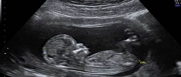
This image shows a baby’s face and hands at 20 weeks, and gives you an idea of what you will be able to see at this scan.
Your doctor will examine all your baby’s organs and take measurements. She will look at:
- The shape, size and structure of your baby’s head and brain. At this stage, severe brain problems, which happen very rarely, are visible.
- Your baby’s face, to check for a cleft lip. Cleft palates inside a baby’s mouth are hard to see and are not often picked up. Fetal eye sockets, eyeballs, nose and ears are also checked.
- Your baby’s spine, both along its length and in cross-section. This is done to make sure all the bones align and that the skin covers the spine at the back.
- Your baby’s abdominal wall, to make sure it covers all the internal organs at the front.
- Your baby’s heart. The top two chambers (atria) and the bottom two chambers (ventricles) should be equal in size. The valves should open and close with each heartbeat. The doctor will also examine the major veins and arteries which carry blood to and from your baby’s heart.
- Your baby’s stomach. Your baby swallows some of the amniotic fluid that he lies in, which is seen in his stomach as a black bubble.
- Your baby’s kidneys. The doctor will check that your baby has two kidneys, and that urine flows freely into his bladder. If your baby’s bladder is empty, it should fill up during the scan and be easy to see. Your baby has been passing urine every half an hour or so for some months now!
- Your baby’s arms, legs, hands and feet. The doctor will look at your baby’s fingers and toes.
- Major and minor soft markers for some common chromosomal abnormalities are also checked.
In addition to this detailed look at how your baby is growing, the doctor will check:
- the placenta
- the umbilical cord
- the amniotic fluid
The placenta may be on the front wall (anterior) or the back wall of your uterus (posterior), usually near the top (or fundus). If the placenta is near the top, it may be described as fundal on your scan report.
The placenta will be described as low if it reaches down to or covers the neck of your uterus (cervix). If the placenta is lying low in your uterus, you’ll have another scan in the third trimester to check its position. By then, it’s likely the placenta will have moved away from your cervix.
Dr. Rajeshwari Pawar
Services including pregnancy care, fetal care, pregnancy sonography and laboratory investigations